Glenohumeral Osteoplasty Augmented Therapy
G.O.A.T.
A novel modification of comprehensive arthroscopic management, incorporating biological augmentation for the arthritic, contracted shoulder — pairing a defined mechanical foundation with an investigational biological construct.
Arthroscopic
Joint preservation
3 layers
Biological adjuncts
≥2 years
Future evaluation
Joint-Preserving Rationale
A mechanical foundation, paired with an investigational biological construct.
Glenohumeral osteoarthritis causes cartilage loss, osteophyte formation, pain and contracture. Arthroplasty is reliable for end-stage disease, but selected younger patients may prioritise native-joint preservation and delay of prosthetic reconstruction. G.O.A.T. combines a defined mechanical foundation with an investigational biological construct.
Restore clearance and movement
- Capsular release to restore clearance and address contracture
- Controlled inferomedial humeral osteoplasty
- Removal of the inferomedial “goat’s beard” osteophyte
- Chondroplasty and treatment of associated intra-articular pathology
Support the treated environment
- Subchondral marrow activation (microfracture)
- A resorbable collagen scaffold
- Autologous micro-fragmented adipose tissue (mFAT)
- Added as biologically plausible adjuncts — not yet validated
Evidence boundary
The mechanical framework is supported by comprehensive arthroscopic management (CAM) and osteoplasty literature. The three-layer biological construct remains investigational and requires prospective clinical validation.
The Preservable Arthritic Shoulder
Proposed patient selection.
Residual joint space, preserved congruity and rehabilitation readiness define the joint-preserving intent.
Included
- Age 35–60 years
- Symptomatic primary or secondary glenohumeral osteoarthritis
- Persistent symptoms after at least six months of structured non-operative care
- Minimum 2 mm joint space and preserved joint congruity
- Ability to comply with structured rehabilitation
Excluded
- Joint space below 2 mm or structurally non-preservable disease
- Advanced posterior glenoid erosion or static posterior subluxation
- Severe rotator-cuff tear arthropathy
- Active infection or uncontrolled inflammatory arthropathy
- Medical factors that preclude safe anaesthesia or rehabilitation
Protocol requirement: prespecify eligibility, imaging thresholds and the handling of borderline glenoid morphology.
Controlled Decompression & Augmentation
A reproducible operative pathway.
Seven defined stages take the procedure from positioning to biological closure, each intended to restore clearance while preserving bone.
Position & inspect
Beach-chair positioning, examination under anaesthesia and systematic glenohumeral inspection.
Create access
Standard posterior and accessory posterior portals establish the inferomedial trajectory.
Release the capsule
Stepwise anterior, posterior and inferior release restores clearance and addresses contracture.
Reshape the humerus
Controlled osteoplasty removes the inferior osteophyte and restores a smooth humeral contour.
Activate marrow
Microfracture recruits marrow-derived repair elements into prepared chondral defects.
Apply the scaffold
A resorbable collagen matrix supports local retention at low flow or during dry arthroscopy.
Add mFAT
Autologous micro-fragmented adipose tissue is introduced under visualisation immediately before closure.
Critical safety principle
Protect the axillary nerve: visualise portal placement, keep the burr on bone and do not direct it below the humeral neck.
Adjuncts to the Mechanical Framework
Three biological layers — clearly bounded.
The biological elements are intended to support the treated environment; the combined shoulder construct is not yet clinically validated.
Marrow activation
Microfracture perforations recruit marrow elements into prepared full-thickness chondral defects.
Evidence status
Established concept; combined role unproven
Collagen scaffold
A temporary resorbable matrix supports local retention of biological material at the treated surface.
Evidence status
Evidence extrapolated from other joints
Micro-fragmented adipose tissue
Autologous mFAT is introduced for its proposed paracrine, immunomodulatory and stromal support.
Evidence status
Emerging, predominantly lower-level evidence
Interpretation
Biological plausibility does not establish clinical efficacy, cartilage restoration, durability or synergy. These outcomes must be tested prospectively.
Claims that should be avoided
Regeneration
Do not present the combined construct as proven cartilage regeneration.
Efficacy
Do not infer comparative effectiveness from component or observational evidence.
Durability
Do not claim long-term joint survival before adequate follow-up is available.
De-Identified Imaging · Mechanical Target
From an inferomedial osteophyte to a restored humeral contour.
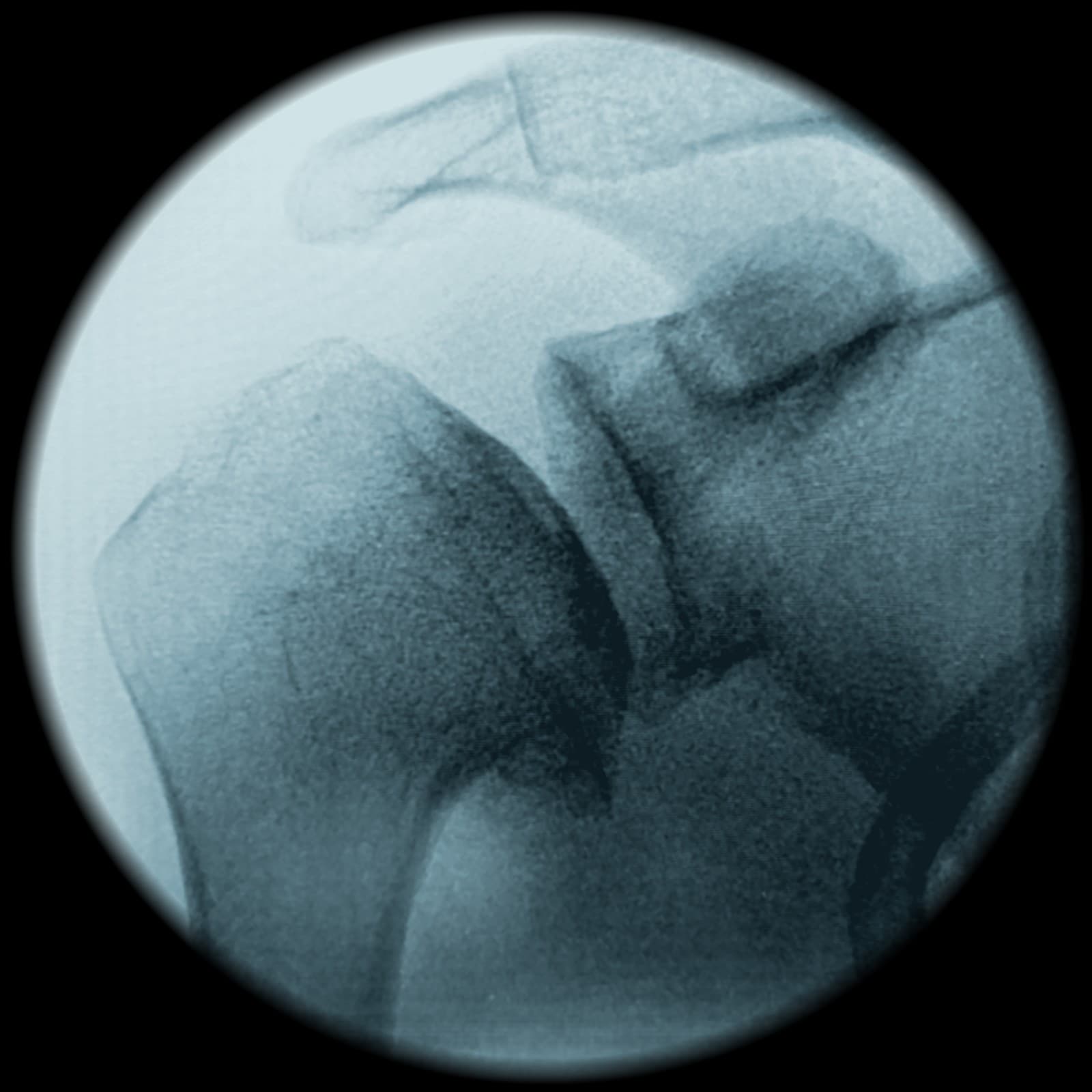
Controlled osteoplasty removes the inferior prominence — the “goat’s beard” osteophyte — and restores a smooth humeral contour.
Before
Inferomedial osteophyte
After
Restored humeral contour
Operative principle: incremental resection to restore clearance while preserving bone and protecting the axillary nerve.
G.O.A.T.
Glenohumeral Osteoplasty Augmented Therapy: a novel modification of comprehensive arthroscopic management incorporating biological augmentation.
Background
Glenohumeral osteoarthritis is progressive and debilitating. G.O.A.T. is described as a modified arthroscopic joint-preserving technique for selected younger or high-demand patients who may prioritise preservation of the native joint and delay of prosthetic reconstruction.
Methods
The procedure uses beach-chair positioning, standard and accessory posterior portals, glenohumeral inspection, capsular release, controlled inferomedial osteoplasty, microfracture of full-thickness defects, collagen-scaffold application and intra-articular mFAT at closure.
Conclusion
G.O.A.T. is a biologically augmented evolution of arthroscopic joint preservation. The mechanical basis is supported by existing literature; the biological construct is an emerging adjunct. Prospective studies are required to establish safety, reproducibility and outcomes.
Study status
Technique description. No clinical results are reported. The combined procedure requires prospective validation.
Evidence Context & Research Agenda
Honest context, deliberate next steps.
~63%
10-year CAM survivorship
CAM evidence; not G.O.A.T. validation
4–42.4%
Conversion range
Heterogeneous arthroscopy studies
36 months
mFAT observation
Injection evidence only
Prospective evaluation priorities
Recruitment
Consecutive enrolment with clearly defined, prospectively applied indications and exclusions.
Outcomes
Standardised safety, pain, function, range-of-motion and conversion-to-arthroplasty reporting.
Follow-up
Minimum two-year follow-up with transparent attrition reporting and predefined analysis.
Comparison
Future comparative work to separate mechanical benefit from any biological contribution.